Longevity Weekly Review 2026-09-22

Week In Review
The dominant theme this week was measurement. For decades the field’s central bottleneck has been that the only definitive test of an anti-aging intervention is a lifespan study, which in mice takes three to four years and in humans is impractical. Three items attack that bottleneck from different angles. A Frontiers in Science paper from Richard Miller, Steven Austad and colleagues proposes twelve “aging rate indicators” that could be read from a single time point and might shrink mouse drug screening to a quarter of the time and a twentieth of the cost. A Nature Aging paper introduces RamanOmics, a label-free imaging method that identifies senescent cells in intact tissue without destroying the sample. And Insilico Medicine, with the Buck Institute and Harvard collaborators, published an open AI toolkit for aging research on the cover of Cell, including a benchmark, a family of small specialized language models, and an autonomous agent that nominated hundreds of candidate targets. Together these point toward a field that is industrializing its tools rather than waiting on serendipity.
A second cluster concerns the immune system and cellular housekeeping as levers on aging. Chinese researchers showed that thymus tissue grafted into the spleen grows far better than in muscle and restores functional T-cell immunity in mice. Stanford scientists found that transplanted bone marrow cells donate healthy mitochondria to neighboring cells in the brain and heart, a mechanism that could extend blood-cell therapies to non-blood organs. A related study in Aging Cell showed that stem-cell secretions protect skin from sun damage by triggering mitophagy, the clearance of damaged mitochondria, and that the benefit disappears when that clearance is blocked. The recurring message is that aging tissues often retain the machinery to repair themselves if given the right cells or signals.
The third theme is that functional decline is more reversible, and more plastic, than the standard narrative suggests. University of Missouri researchers identified a failure of nerve-to-muscle signaling as a driver of age-related weakness and reversed it in old rodents with a drug already in human trials. An Oregon Health & Science University pilot found that time-restricted eating improved a neurodegeneration biomarker in early Huntington’s disease. University of Arizona psychologists argued that the aging brain is adapting rather than declining, shifting from detail capture to meaning-making. And a sober review in The Conversation asked whether dietary calorie-restriction mimetics actually deliver in humans, concluding that the evidence still favors diet quality and exercise over any single compound.
Read together, the week suggests a field maturing in both ambition and discipline. Faster and cheaper measurement makes it possible to test many more interventions. Immune and mitochondrial repair strategies offer concrete candidates to test. And the human-facing studies, from muscle to brain to diet, are increasingly careful to distinguish promising signals from proven effects.
Items
Twelve Aging Rate Indicators Could Cut Anti-Aging Drug Screening to a Quarter of the Time
The single biggest practical obstacle in aging research is time. To learn whether a drug slows aging in mice, researchers must wait for animals to die, which takes three to four years and costs a great deal of money. Testing in humans is even less feasible. A new article in Frontiers in Science, led by Richard Miller of the University of Michigan and Steven Austad of the University of Alabama at Birmingham, proposes a way around this constraint.
The authors describe twelve candidate “aging rate indicators,” measurable biological signals that could reveal whether an intervention is slowing aging from a single measurement at one time point. This contrasts with conventional biological age tests, which typically require repeated measurements over long periods. The indicators were drawn from analysis of fourteen agents or drug combinations already proven to extend mouse lifespan, eight of which produced lifespan increases of twelve percent or more.
The authors estimate that if the indicators can reliably distinguish effective drugs from ineffective ones, mouse studies could be completed roughly four times faster and at about one-twentieth the cost. That would let the field screen many more candidates before committing to expensive full lifespan trials. Miller noted that the indicators “may allow us to see which drugs can induce patterns more similar to slow aging mice.” Austad added that many effective anti-aging drugs “remain fully active even when started in late adulthood,” a point with obvious relevance to human use.
The proposal comes with caveats. The indicators have been derived from mouse data and require validation across other mammalian species and eventually in humans before they can guide clinical decisions. But as a framework, it addresses the field’s most persistent logistical problem head-on.
Source: EurekAlert
Insilico Publishes an Open AI Toolkit for Aging Research on the Cover of Cell
Insilico Medicine, working with Liquid AI, the Buck Institute for Research on Aging, Harvard Medical School and Brigham and Women’s Hospital, published a study in Cell that was selected as the cover feature of the journal’s September 17 issue. The paper introduces three interconnected open-source resources intended to accelerate AI-driven aging research worldwide.
The first is LongevityBench, described as the first open benchmark for evaluating AI reasoning across five domains of aging biology: clinical data, genetics, epigenetics, transcriptomics and proteomics. The second is a family of five compact “Longevity-LLMs” ranging from 0.6 to 9 billion parameters, fine-tuned on aging-specific clinical and multi-omics data. The third is Longevity Claw, an autonomous research platform that combines the specialized models with scientific tools for gene enrichment analysis, aging-clock calculation and target prioritization.
The benchmark results are striking. The team evaluated eighteen frontier AI systems from OpenAI, Google, Anthropic, xAI, DeepSeek and Moonshot AI, and found that no single system dominated across all biological domains. The best-performing specialized model, a 9-billion-parameter variant built on Qwen, outperformed all twenty-six tested systems. Even the smallest 0.6-billion-parameter model exceeded most frontier systems on these aging-specific tasks. Longevity Claw nominated 328 potential aging intervention targets with up to 5.6-fold enrichment against validated targets, and one nominated gene, KDM1A, was independently validated as extending lifespan in the roundworm C. elegans.
Founder and co-CEO Alex Zhavoronkov said the longevity community “is moving beyond static aging clocks toward foundation models capable of generating measurable, actionable insights.” The models, code and a public leaderboard are available openly, which matters: the value of a benchmark comes from others using it.
Source: EurekAlert
RamanOmics Identifies Senescent Cells in Intact Tissue Without Destroying the Sample
Senescent cells, the damaged cells that stop dividing but linger and secrete inflammatory signals, are a central target in aging research. Yet identifying them in tissue has typically required destructive preparation and molecular labels. A paper published in Nature Aging on September 21 describes a platform called RamanOmics that combines label-free hyperspectral Raman imaging with single-nucleus RNA sequencing and spatial transcriptomics to profile senescence at single-cell resolution.
Raman imaging reads the vibrational signatures of molecules inside cells, so it can characterize chemical composition without staining. By pairing this with sequencing data from the same tissue, the researchers built a machine-learning-derived multimodal “barcode” that identifies senescent cells in place, nondestructively.
The findings show that senescence is tissue-specific. In lung, senescent cells were enriched for extracellular matrix remodeling and TGF-beta signaling. In skin, they expressed epidermal differentiation genes. Across tissues, however, a conserved lipid-associated Raman signature marked cells expressing the senescence marker p21. The team validated the approach in wound healing, where senescent cells reactivated epidermal differentiation programs alongside stronger lipid signatures.
The practical significance is that senescence could become something to watch in living tissue over time rather than something to infer from destroyed samples. That capability would help both basic research and the evaluation of senolytic drugs, complementing the aging rate indicators proposed elsewhere this week.
Source: Nature Aging
Thymus Grafts Grow in Mouse Spleens and Restore Immunity
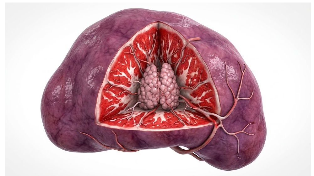
The thymus, where T cells mature, shrinks dramatically with age, and its decline is a major reason older people respond poorly to infections and vaccines. Regenerating or replacing thymic function is therefore a long-standing goal. A Chinese research group reporting in Advanced Science found that where you put a thymus graft matters enormously.
The team compared thymic tissue transplanted into the spleen against the conventional site, muscle. Splenic grafts grew substantially faster and reached roughly half of normal thymus weight, whereas muscle grafts were smaller and less organized. By week two, splenic grafts had developed the organized cortical and medullary regions characteristic of a functioning thymus. T-cell counts improved, though CD4-positive cells stabilized at about half normal levels.
Function followed structure. Mice with splenic grafts controlled vesicular stomatitis virus infection better than untreated controls. Against B16 melanoma, graft recipients showed restricted tumor growth with more tumor-infiltrating T cells. When challenged with human colorectal cancer cells, splenic-graft recipients rejected the tumors entirely, matching immunocompetent controls. The researchers attribute the spleen’s advantage to its rich blood supply, its immune-tolerant environment and its secretion of growth factors. Experiments with human tissue suggested the approach could translate.
The result is a reminder that regenerative medicine depends as much on the niche as on the tissue. It also offers a concrete route toward rebuilding adaptive immunity in aged individuals.
Source: Lifespan.io
Transplanted Immune Cells Donate Their Mitochondria to Failing Neighbors
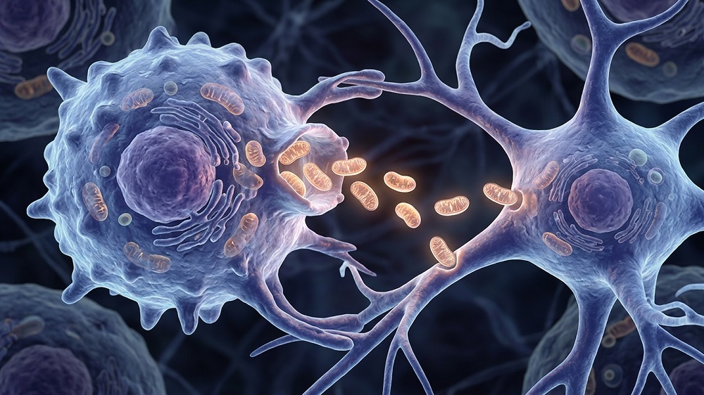
Bone marrow transplants replace the blood and immune system. A Stanford University study in Nature Communications shows they can do something less expected: deliver working mitochondria to cells in organs far from the blood. The work, led by Natalia Gomez-Ospina, used a mouse model of Friedreich’s ataxia, an inherited mitochondrial disease.
After transplanting healthy bone marrow cells and tracking them with fluorescent labels, the researchers found that donor-derived immune cells took up residence throughout the body. About 82 percent of brain microglia were donor-derived five months after transplantation. Those cells then handed off their mitochondria to neighboring cells whose own mitochondria were failing, including neurons and supporting cells in the brain and heart muscle cells. About 22 percent of recipient cells in bone marrow showed mitochondrial signals from donors, and transfer required direct cell-to-cell contact.
The functional benefits were meaningful. Female survival improved from 53 percent to 80 percent with healthy marrow treatment. Treated mice recovered roughly half of their weight deficit and improved on tests of coordination, spontaneous movement and strength, though not to healthy levels. Molecular analysis showed increased expression of genes for oxidative phosphorylation and ATP synthesis in recipient tissues.
Gomez-Ospina described it as “a way to use the blood system to treat nonblood organs,” and noted that “cells are talking to each other in ways that are more consequential than we’ve realized.” Mitochondrial dysfunction is a hallmark of aging as well as of inherited disease, so a mechanism for delivering fresh mitochondria to tissue has implications well beyond Friedreich’s ataxia.
Source: Lifespan.io
Clearing Damaged Mitochondria Protects Skin From Sun-Driven Aging
Ultraviolet exposure is the largest external driver of skin aging, and current treatments such as retinoids offer only partial protection. A study in Aging Cell reports that secretions from human umbilical cord mesenchymal stem cells can reduce sun-induced damage in mouse skin and human skin cells, and identifies the mechanism.
Mice exposed to 40 days of UVA and UVB radiation and treated with the stem cell secretome showed less epidermal thickening, better hydration, improved elasticity and less water loss than untreated animals. Collagen largely remained intact and markers of cellular senescence fell significantly. Human skin cells treated the same way showed reduced senescence and preserved structural proteins.
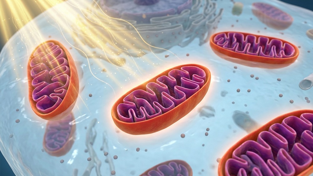
The key finding is how it works. The secretome triggered mitophagy, the process by which cells clear damaged mitochondria. That clearance suppressed inflammatory signaling through the cGAS-STING pathway, which is activated when mitochondrial DNA leaks into the cell. When the researchers blocked mitophagy, the protective effect vanished, confirming that mitochondrial cleanup was essential rather than incidental.
The result connects skin aging to the same mitochondrial quality-control and innate-immune pathways implicated in aging elsewhere in the body. The next steps are to isolate the active compounds in the secretome and test them in humans, since the current work relies on mouse models and immortalized cell lines.
Source: Lifespan.io
Time-Restricted Eating Shows Promise in an Early Huntington’s Disease Trial

Intermittent fasting is among the most studied lifestyle interventions in aging research, with strong animal data and mixed human results. A pilot trial from Oregon Health & Science University, published in Nature Metabolism, tested time-restricted eating in twenty people with early-stage Huntington’s disease, a neurodegenerative condition with a strong metabolic component.
Participants, with an average age of 45 and evenly split by sex, adopted a six-to-eight-hour daily eating window for twelve weeks. Most chose windows starting between 10 a.m. and 1 p.m. and ending between 6 p.m. and 8 p.m. Adherence was good and weight remained stable, with an average loss of about 1.1 kilograms.
The clinical signals were encouraging for a pilot. The Unified Huntington’s Disease Rating Scale improved by half a point, against a typical annual decline of roughly one point. Plasma neurofilament light, a blood marker of neuronal damage that normally rises as the disease progresses, fell by a mean of 12.6 percent. Blood cells isolated from participants showed enhanced oxygen consumption and increased ATP production, suggesting a metabolic effect at the cellular level. Participants also reported about 37 more minutes of physical activity per week.
Lead researcher Russell Wells called the findings “remarkable” for a pilot study, while acknowledging that the lack of a control group and concurrent lifestyle changes limit what can be concluded about time-restricted eating on its own. A controlled trial is the obvious next step. For the longevity field, the study adds a human data point that fasting-based interventions can shift neurodegeneration biomarkers, not just metabolic ones.
Source: Lifespan.io
Age-Related Muscle Weakness Traced to Failing Nerve-Muscle Communication, and Reversed in Animals
Sarcopenia, the loss of muscle strength with age, affects nearly half of adults over 80 and is a leading cause of falls and lost independence. The prevailing view has been that it results primarily from loss of muscle mass. A University of Missouri-led study in the Journal of Clinical Investigation identifies a different, and potentially reversible, cause.
The team, led by W. David Arnold with international collaborators from Denmark, Scotland, Saudi Arabia, India and Japan, found that in weak older individuals the neuromuscular junction, the point where nerves signal muscles to contract, fails to transmit reliably. The degree of transmission failure correlated with the severity of weakness. Looking closer, they found a localized reduction of the sodium channel NaV1.4 at the muscle side of the junction, which makes aging muscle less responsive to nerve signals.
Crucially, the defect could be corrected. Partial inhibition of the chloride channel ClC-1 enhanced muscle excitability and improved both neuromuscular transmission and muscle function in old rodents. “The neuromuscular junction is failing with aging,” Arnold said, but the discovery shows this failure “is potentially reversible.”
The translational path is unusually short. A ClC-1 inhibitor called ignaseclant, developed by NMD Pharma, has already shown improvements in muscle strength measures in patients with Charcot-Marie-Tooth disease, according to trial results presented at the 2026 Muscular Dystrophy Association conference. The researchers are optimistic it could eventually help older adults with sarcopenia, which would make it one of the first drugs to target an age-related functional decline directly.
Source: University of Missouri
The Aging Brain Is Not Declining. It Is Adapting, Researchers Argue
A framework paper in Perspectives on Psychological Science challenges the assumption that cognitive aging is simply decline. Fabian-Xosé Fernandez of the University of Arizona, with coauthors Sara Burke and Lynn Nadel, proposes what they call the adaptive aging hypothesis: that late-life cognition is a distinct, evolutionarily honed phase of human development that prioritizes wisdom and the sharing of stored expertise over the capture of fine detail.
The argument draws on molecular biology and neuroimaging. In healthy aging, activity in the hippocampus, which handles precise episodic details, decreases, while activity in the frontal cortex, which processes general concepts and meaning, increases. The authors interpret this shift not as compensation for damage but as purposeful maturation, suited to mentoring, judgment and the transfer of knowledge across generations. “The older brain is optimized for different things than the younger brain,” Fernandez said, “predominantly the sharing of stored expertise.”
The hypothesis also offers a way to distinguish normal aging from disease. In Alzheimer’s disease, Fernandez notes, “the hippocampus is hyperactive,” preventing the expected transition. That distinction could sharpen diagnostic thinking about what is pathological and what is not.
The practical implication is that rather than trying to force aging brains to retain granular details, society should leverage older adults’ strengths in wisdom-sharing and intergenerational teaching. For a field focused on extending healthspan, the paper is a useful reminder that the goal is not to freeze people at 30, but to support the capabilities each stage of life is built for.
Source: Association for Psychological Science
Can Nutrients in Food Mimic Calorie Restriction? The Human Evidence, Weighed
Calorie restriction reliably extends lifespan in laboratory animals, but few people can sustain it. That has fueled interest in “calorie-restriction mimetics,” compounds found in ordinary foods that trigger similar cellular pathways: reducing growth signals, encouraging autophagy, and influencing how cells sense energy and support their mitochondria. Justin Roberts, a professor of nutritional physiology, reviews the human evidence in a piece for The Conversation.
The headline conclusion is cautionary. While calorie-restriction mimetics show promise in cellular and animal studies, current human evidence does not demonstrate that they reproduce the effects of calorie restriction or extend healthspan. The CALERIE trial, which followed 218 adults for two years, showed that calorie restriction itself improved cardiovascular and metabolic markers but could not establish lifespan extension.
The specific compounds have mixed records. Spermidine, found in wheat germ, mushrooms and legumes, has been linked to lower mortality in observational studies, but a twelve-month randomized trial found no memory improvement versus placebo. Urolithin A improved muscle endurance and mitochondrial measures but did not significantly improve walking distance or peak muscle power. Meanwhile, a 2025 study of about 105,000 adults found that healthy dietary patterns rich in fruits, vegetables, whole grains, nuts and legumes were associated with aging free of chronic disease.
Roberts’s recommendation is that “the strongest evidence for aging well supports a varied, nutritious diet and purposeful physical activity.” The piece is a healthy counterweight in a week full of promising mechanisms: in humans, the well-established basics still outperform any single molecule.
Source: The Conversation